What Is a CT Machine and How Does It Work?
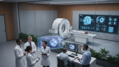
A Ct Machine is more than a large imaging device. It is a carefully engineered system that combines rotating X-ray tubes, detectors, computers, and reconstruction software. Unlike a standard radiograph, it collects multiple measurements around the body. The computer then converts those measurements into cross-sectional images, often called slices. These slices can reveal bleeding, fractures, tumors, infection, and internal damage within seconds.
The technology began with a bold idea. Nobel Prize-winning engineer Godfrey Hounsfield described it this way: “I had a vague idea that you could use a computer to reconstruct an image of the inside of the body.” His insight changed diagnostic medicine. Today, modern scanners can create thin images of the chest while patients remain still for only a few breaths. The result feels simple. The engineering is not.
NCRP Report No. 160 estimated that CT represented about 24% of diagnostic radiological examinations but contributed approximately 67% of the collective effective dose in the United States. That finding explains why image quality and radiation control must develop together. The IAEA also emphasizes justification and optimization for medical exposure. In practice, technologists adjust tube current, voltage, scan length, and reconstruction settings for each patient. A smaller child should not receive an adult protocol.
This article explains what happens inside a Ct Machine, from X-ray production to digital image reconstruction. It also examines contrast media, safety checks, scan limitations, and common misconceptions. Some explanations remain simplified. Real clinical decisions depend on patient history, equipment performance, and professional judgment. That complexity deserves attention.
What Is a CT Machine?
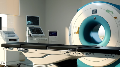
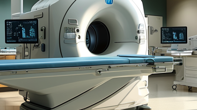
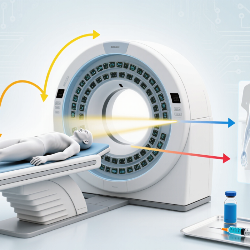
A CT machine is a medical imaging system that creates detailed cross-sectional pictures of the body. CT stands for computed tomography. Unlike a standard X-ray, it shows tissues from multiple angles. It is not an ordinary X-ray. A large, ring-shaped scanner surrounds the patient while an X-ray tube rotates inside it. Detectors measure how much radiation passes through the body. A computer then converts these measurements into thin image slices, which can be viewed individually or combined into a three-dimensional model.
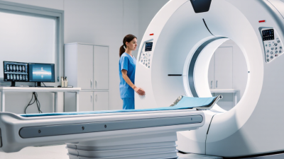
The patient usually lies on a narrow table that moves through the scanner’s opening. The process is generally quick and painless, although staying still is essential for clear images. In some examinations, a healthcare professional may use contrast material to make blood vessels or certain organs easier to see. This can create a warm sensation or a brief metallic taste. CT scans use more radiation than ordinary X-rays, so trained clinicians request them when the expected medical value justifies the exposure. Modern systems can produce excellent detail, but they are not perfect. Motion, metal objects, or unclear clinical questions may reduce accuracy. The final interpretation should come from a qualified radiologist alongside the patient’s symptoms and other test results. A picture helps. It does not tell the whole story.
What Is a CT Machine and How Does It Work?
A computed tomography (CT) machine uses a rotating X-ray tube and detectors to collect measurements from multiple angles. A computer reconstructs these measurements into cross-sectional images. The chart shows representative Hounsfield Unit values, a scale used to describe how different materials attenuate X-rays in CT images.
Water is defined as approximately 0 HU. Values are representative reference points; actual measurements can vary with tissue composition, scan settings, and reconstruction methods.
How Does a CT Scan Work Step by Step?
A CT machine combines X-rays with computer processing to create cross-sectional images of the body. The scanner looks like a short, open tunnel. Inside it, an X-ray tube rotates around the patient while detectors measure the passing radiation. A computer then converts these measurements into thin “slices,” which can be viewed individually or as a three-dimensional model.
The process begins when the patient lies on a narrow table. A technologist checks the requested body area and removes nearby metal objects. The table moves slowly through the scanner’s opening. The machine may take a quick planning image before the detailed scan starts. In some examinations, a contrast solution is given by mouth or through a vein. It can make blood vessels, organs, or abnormal tissue easier to see.
During image collection, the patient may hear brief buzzing or clicking sounds. The technologist often asks for a few seconds of stillness or breath-holding. Small movements can blur the images, so this instruction matters. The X-ray tube then circles the body, collecting information from many angles. Software reconstructs those readings into images that a radiologist reviews.
A CT scan is fast, but it is not a perfect photograph. Image quality can change with movement, metal, body position, or limited contrast. Radiation exposure is carefully considered, especially for children and pregnancy. Patients should report allergies, kidney problems, and possible pregnancy before scanning.
How Are CT Images Created and Interpreted?
A CT machine creates images by combining many X-ray measurements. An X-ray tube rotates around the patient. Detectors record how much radiation passes through the body. Dense tissues absorb more radiation and appear brighter. Air appears darker.
A computer converts these measurements into thin cross-sectional slices. It then reconstructs them into views from different angles. The result resembles a detailed map of internal structures, not a normal photograph.
Technologists position the patient carefully and check for movement. Even slight motion can blur small findings. Contrast material may highlight blood vessels, organs, or areas of inflammation. It must be used thoughtfully, especially when kidney function or allergies are concerns.
Radiologists interpret CT images using clinical information, prior scans, and several display settings. One setting may emphasize lung detail. Another may reveal subtle changes in soft tissue. They examine patterns, shapes, symmetry, and measurements rather than isolated bright spots. The process is not flawless. Metal implants, motion, and unusual anatomy can create artifacts. A harmless finding may also look concerning at first glance. Experienced interpretation therefore includes comparison, uncertainty, and careful wording. CT images support diagnosis, but they do not replace physical examination or professional judgment.
What Are the Main Uses and Safety Considerations of CT Scans?
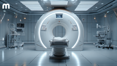
A CT machine combines X-rays with computer processing to create detailed cross-sectional images. The scanner looks like a short tunnel with a moving table. As the table passes through, an X-ray tube rotates around the body. A computer then builds slices that doctors can review individually or as three-dimensional images.
CT scans help investigate head injuries, chest pain, stroke symptoms, kidney stones, internal bleeding, and suspected tumors. They can also guide biopsies and monitor treatment. However, CT is not automatically the best test. It uses ionizing radiation, so clinicians should consider the patient’s age, symptoms, medical history, and previous imaging. Children and pregnant patients may need special assessment. The image is powerful, but not perfect. Small findings can still be unclear.
Tips:
Tell the imaging team about pregnancy, allergies, kidney disease, diabetes, or implanted devices. Ask whether contrast material is necessary and what side effects require attention. Wear comfortable clothing and follow instructions about food or drink. During the scan, remain still and breathe as directed. The procedure is usually quick and painless. After contrast, some people notice brief warmth or a metallic taste. Serious reactions are uncommon, but urgent symptoms need immediate medical attention. Ask how the result will be explained; uncertainty can remain until a qualified radiologist reviews the images.