What Is the Best Medical CT Scan for Hospitals?

Choosing the best Medical Ct Scan for a hospital is not simply a purchasing decision. It affects diagnosis, radiation exposure, workflow, and long-term operating costs. The World Health Organization and International Atomic Energy Agency both emphasize equipment quality, trained staff, protocol control, and patient safety. Their guidance makes one point clear: higher image quality alone does not define the best scanner.

Recent industry analyses from IMV and market research firms show strong hospital demand for 64-slice, 128-slice, and advanced spectral CT systems. However, market popularity can mislead. A rural hospital may need reliability, low maintenance costs, and strong emergency imaging. A tertiary center may require cardiac CT, dual-energy imaging, and rapid trauma protocols. The room matters too. A scanner that looks impressive on paper may struggle beside a crowded emergency department.

Dr. Cynthia McCollough, a Mayo Clinic medical physicist and CT dose expert, has repeatedly promoted a practical principle: “The best CT protocol is the one that answers the clinical question with the lowest reasonable dose.” That principle should guide equipment selection. Not the largest number of slices.

A credible comparison must examine detector coverage, iterative reconstruction, dose monitoring, uptime, service response, cybersecurity, and staff training. The lowest purchase price may become expensive after repeated delays. Yet specifications do not tell the whole story. Hospitals should review local patient volume, radiologist expertise, and real-world test scans before choosing. Mistakes happen. Careful evaluation remains safer than following a brochure.
What Is the Best Medical CT Scan for Hospitals?

Define the Best Hospital CT by Clinical Need, Dose, Speed, and Total Cost

What Is the Best Medical CT Scan for Hospitals?

The best hospital CT scanner depends on clinical need, not a single technical score. In an emergency department, rapid image acquisition can support stroke, trauma, and pulmonary embolism pathways. Speed matters. However, fast scanning should not replace image quality or careful patient positioning.

Dose management remains essential, especially for children, repeat examinations, and patients needing regular follow-up. A suitable system should adjust exposure to body size and examination type. Radiologists, technologists, and medical physicists should review protocols together. They can compare diagnostic quality, noise levels, and dose reports using local patient data. Numbers can mislead. A lower dose is not useful if doctors cannot interpret the images confidently.

Total cost also extends beyond the purchase price. Hospitals should assess contrast delivery, room preparation, staff training, maintenance, software updates, energy use, and possible downtime. A scanner that completes more examinations per hour may improve access, but only when staffing and reporting capacity match that speed. Reliability deserves attention. One missed service day can disrupt referrals and delay treatment. Clinical teams should test representative cases before selection, including obese patients, motion-prone patients, and complex contrast studies. The perfect specification may look impressive, yet everyday workflow can reveal weaknesses.
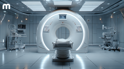
Compare 16-, 64-, 128-, and 256-Slice CT Detector Configurations

What Is the Best Medical CT Scan for Hospitals?

The best CT system depends on clinical workload, not detector count alone. A 16-slice scanner can handle routine head, chest, abdominal, and trauma examinations efficiently. It usually costs less and demands simpler infrastructure. However, slower coverage may affect obese patients, breath-hold studies, and emergency imaging.

A 64-slice configuration offers a practical balance for many hospitals. It supports faster angiography, multiphase studies, and broader emergency coverage. A 128-slice system can improve temporal resolution and cardiac imaging when protocols and ECG control are properly managed. A 256-slice detector covers a larger anatomical range per rotation. That can reduce motion during pediatric, perfusion, and dynamic cardiac examinations. Coverage varies by detector width and pitch, however. Slice labels can mislead.

The American College of Radiology Dose Index Registry reports CTDIvol and DLP benchmarks across large examination datasets. These metrics show why protocol optimization matters more than headline slice numbers. The International Atomic Energy Agency recommends local diagnostic reference levels, commonly based on the 75th percentile of dose distributions. Hospitals should compare image quality, scan time, repeat rates, uptime, and dose. A 256-slice system is not automatically safer. A well-calibrated 64-slice unit may perform better in a disciplined department. I would also question whether every hospital needs advanced cardiac capability. That investment can become underused capacity.

Benchmark Radiation Using FDA’s Typical 1–10 mSv CT Dose Range

What Is the Best Medical CT Scan for Hospitals?

A hospital’s best CT scan is not simply the machine with the lowest radiation number. The right choice depends on the clinical question, image quality, patient size, and available dose controls. FDA’s typical 1–10 mSv CT dose range offers a practical benchmark, not a promise for every examination.

A head CT may sit near one part of that range, while chest, abdominal, or multiphase studies can be higher. Actual exposure changes with protocol, anatomy, scan length, and repeated contrast phases. Dose matters. So does diagnostic confidence. A scan that uses less radiation but misses a small finding may create another clinical problem.

In practice, hospital teams should review the expected dose and the reason for each scan series. Dose reports commonly include CTDIvol and DLP, which help professionals compare protocols, but they are not a patient’s exact risk. Children, smaller adults, and patients needing several scans deserve extra attention. The 1–10 mSv benchmark is useful, yet imperfect. “Low dose” can sound reassuring, while excessive reduction may weaken useful detail. That trade-off deserves honest discussion between radiologists, technologists, and referring clinicians.

What Is the Best Medical CT Scan for Hospitals? - Benchmark Radiation Using FDA’s Typical 1–10 mSv CT Dose Range

A practical comparison of common hospital CT protocols. The “best” examination depends on the clinical question, patient characteristics, image-quality requirements, and the need for contrast or multiphase imaging.

CT Protocol Typical Clinical Use Approximate Effective Dose Position Relative to FDA’s Typical 1–10 mSv Range Diagnostic Strength Main Limitations Hospital Selection Consideration
Low-dose chest CT Lung cancer screening and selected follow-up of pulmonary nodules About 1–2 mSv At or near the lower end Good detection of small pulmonary nodules and many early lung abnormalities Limited evaluation of vascular structures, mediastinum, and some non-lung conditions Useful when a validated low-dose protocol and appropriate patient-selection pathway are available
Routine chest CT Infection, malignancy, interstitial lung disease, trauma, and mediastinal assessment About 5–7 mSv Within the typical range Detailed assessment of lung parenchyma, pleura, mediastinum, and chest wall Higher dose than screening protocols; intravenous contrast may be required for some indications Strong general-purpose option for emergency and inpatient chest imaging
Non-contrast head CT Acute hemorrhage, head injury, hydrocephalus, and initial stroke assessment About 2–4 mSv Within the typical range Fast detection of acute intracranial hemorrhage, mass effect, and skull injury Less sensitive than MRI for many early ischemic, posterior fossa, and soft-tissue abnormalities Essential emergency capability because of speed, availability, and compatibility with unstable patients
CT angiography of the head and neck Aneurysm, arterial stenosis, large-vessel occlusion, and vascular trauma About 4–7 mSv Within the typical range Rapid visualization of intracranial and cervical vessels after iodinated contrast administration Requires contrast; image quality can be affected by motion, calcification, and poor vascular timing Valuable for hospitals providing comprehensive stroke and vascular emergency services
CT pulmonary angiography Evaluation of suspected pulmonary embolism About 3–6 mSv Within the typical range High diagnostic performance for clinically significant pulmonary embolism Requires iodinated contrast and careful protocol selection; radiation exposure to radiosensitive organs should be considered Best suited to an emergency workflow that includes pre-test probability and laboratory assessment
Routine abdomen and pelvis CT Abdominal pain, infection, obstruction, trauma, and suspected malignancy About 7–10 mSv At the upper end Broad evaluation of abdominal and pelvic organs, bowel, vessels, and retroperitoneum Higher radiation burden; contrast may be needed, and renal function or allergy history may affect planning Important general-purpose inpatient and emergency protocol; dose optimization is especially valuable
CT for suspected renal colic Detection of urinary stones and complications of obstruction About 2–5 mSv with a reduced-dose protocol Low to middle portion Excellent detection of many urinary calculi and alternative causes of acute flank pain Reduced-dose imaging may be less suitable for very large body habitus or complex alternative diagnoses A dedicated reduced-dose pathway can lower exposure while preserving the required diagnostic information
Coronary CT angiography Selected patients with suspected coronary artery disease or anomalous coronary anatomy About 1–5 mSv with modern prospective techniques Low to middle portion Noninvasive assessment of coronary anatomy and plaque in appropriately selected patients Heart-rate control, rhythm stability, contrast timing, and calcium burden affect image quality and interpretation Best where cardiac gating, trained interpretation, and a coordinated cardiology-radiology workflow are available
Multiphase liver or pancreas CT Characterization of focal liver lesions, pancreatic disease, and selected treatment planning About 10–25 mSv, depending on the number of phases Often above the typical single-scan range Dynamic enhancement patterns can improve lesion characterization and staging Multiple acquisitions increase radiation exposure and contrast use; not appropriate when a single-phase study is sufficient Use only when the added phases are clinically justified and protocol timing is optimized
CT colonography Colorectal polyp and cancer evaluation when optical colonoscopy is incomplete or unsuitable About 4–8 mSv Within the typical range Noninvasive evaluation of the colon and selected extracolonic findings Does not permit biopsy or polyp removal; bowel preparation and colonic insufflation are required Useful as an alternative diagnostic pathway, with follow-up colonoscopy required for significant findings
Interpretation: Effective-dose values are approximate population-level estimates, not patient-specific measurements. Actual dose varies with patient size, anatomy, scan length, scanner settings, reconstruction method, contrast phases, and clinical indication. Dose should be kept as low as reasonably achievable while maintaining adequate diagnostic image quality.
Reference basis: U.S. Food and Drug Administration educational guidance describing typical CT effective-dose ranges; published radiology dose estimates commonly used for adult examinations. Effective dose is not intended for direct individual risk prediction.

Use ACR CTDIvol and DLP Benchmarks to Assess Dose Efficiency

What Is the Best Medical CT Scan for Hospitals?

The best hospital CT scan delivers diagnostic images at the lowest reasonable radiation dose. Dose efficiency should be measured, not guessed. ACR CTDIvol and DLP benchmarks provide practical reference points for this review. CTDIvol estimates scanner output during a protocol, while DLP reflects dose across the scanned length. Neither value describes a patient’s exact absorbed dose.

Hospitals should compare similar examinations, body regions, and patient groups. A chest CT may show acceptable CTDIvol but an excessive DLP if the scan range is too long. Patient size also matters. A small adult and a large adult can require different exposure settings. Benchmark values are guides, not automatic pass-or-fail rules. Image quality must remain clinically useful. A lower number is not always better.

Tips: Review dose data by protocol and patient size. Check repeated scans and unusually long ranges. Ask a medical physicist and radiologist to investigate persistent outliers. Document changes, then reassess image quality. The process is rarely perfect. Some protocols need adjustment, and local practice may differ from published benchmarks. That is acceptable when the clinical reason is clear, the decision is documented, and patient safety remains central.

What Is the Best Medical CT Scan for Hospitals?

Comparing routine adult CT protocols with ACR CTDIvol and DLP reference benchmarks helps hospitals assess dose efficiency while maintaining diagnostic image quality.

Lower values generally indicate greater dose efficiency, but protocol selection should also consider patient size, scan range, clinical indication, and image quality. CTDIvol is reported in mGy, while DLP is reported in mGy·cm. Values shown are adult examination reference benchmarks commonly used for CT dose assessment and are not individual patient dose limits.

Verify Cardiac, Trauma, Oncology, Pediatric, and AI Workflow Performance

The best hospital CT scanner should prove performance across real clinical pathways, not merely advertise detector count. Cardiac testing should measure ECG synchronization, temporal resolution, calcium blooming, and motion artifacts. A 2023 European Society of Cardiology guideline supports ECG-gated CT for selected coronary assessments, but local heart-rate control remains important. Trauma testing should record scan-to-report time, whole-body coverage, contrast timing, and dose. Every minute matters.

Oncology protocols require stable low-contrast detectability and repeatable measurements for follow-up imaging. The American College of Radiology’s 2024 Dose Index Registry shows why benchmarking matters: participating facilities compare CT dose indices by examination type, rather than relying on manufacturer claims. Pediatric verification needs age- and size-adjusted protocols, automatic exposure control, and visible dose alerts. Children need more care. ICRP Publication 135 emphasizes diagnostic reference levels as optimization tools, not fixed limits.

AI workflow performance deserves separate validation. The U.S. FDA’s 2024 AI-enabled medical device list contained more than 800 authorized devices across specialties, yet authorization does not guarantee local sensitivity, uptime, or clinical usefulness. Hospitals should measure alert latency, false positives, missed findings, radiologist acceptance, and integration with reporting systems. A 2024 RSNA workflow report also emphasizes human oversight and monitoring after deployment. No protocol is perfect. Scanner selection should include repeat testing, independent physicist review, and difficult cases, because polished demonstrations rarely reveal pediatric motion, metallic implants, or overloaded emergency workflows.